Predicting Residual Neoplasia Risk After “Noncurative” Endoscopic Submucosal Dissection for Malignant Colorectal Lesions
Douglas K. Rex, MD, MASGE, reviewing Santos-Antunes J, et al. Endoscopy 2022 Jul 21.
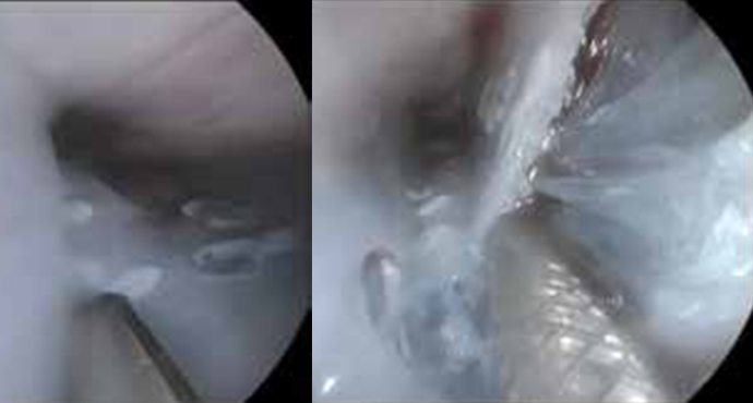
This is a retrospective study from 15 Western centers performing colorectal endoscopic submucosal dissection (ESD) in Europe and Australia.
A curative resection was defined as R0, only mucosal disease or superficial submucosal invasion (SMI <1000 microns), well or moderately differentiated, and without lymphovascular invasion (LVI). All other resections and those presenting with tumor budding were considered noncurative.
Among 2255 colorectal ESDs, there were 381 noncurative resections, of which 352 received either surgery or at least one follow-up endoscopy. Of these, 135 were T1 cancers, including 60 colonic and 75 rectal.
Eight patients refused surgery, 15 had indications for surgery but were too ill, and 96 (71%) underwent surgery. One patient died from surgical complications, and the median endoscopic follow-up of patients who did not undergo surgery was 12 months.
Of the 135 patients with malignant lesions, 17 (13%) had superficial SMI, and none of these patients had lymph node-positive or residual disease in the bowel wall, regardless of other risk factors. The remainder of patients had deeper SMI, and residual disease was discovered in 24% of these patients. However, in the absence of poor differentiation, positive horizontal or vertical margins, and LVI, the risk of lymph node metastasis (LNM) or residual wall lesion was also 0%.
The authors developed a predictive score for LNM. Lymphatic invasion was scored with 2 points, and poor differentiation received 1 point. These were the only two factors that predicted LNM after logistic regression. For scores of 0, 1, 2, and 3, the risk rates of positive lymph nodes were 6%, 25%, 30%, and 75%, respectively.
Risk factors for residual disease in the bowel wall were piecemeal resection, poor differentiation, and positive or uncertain vertical margin. Based on multivariate analysis, a score of 1 was assigned to poor differentiation and a score of 2 was assigned for piecemeal resection, involved vertical margin, or unknown vertical margin. For scores of 0, 1 to 3, and >3, the risk of residual disease in the bowel wall was 2%, 18%, and 50%, respectively.

COMMENTMost patients with cancer in EMR or ESD specimens will have negative surgical resections. There are increasing efforts to provide a more quantitative assessment of the risk of residual disease. For both EMR- and ESD-resected malignant polyps, recent work shows that LNM is nearly always negative in the absence of poor differentiation and LVI. Further, residual disease in the bowel wall is related to positive vertical margins after both EMR and ESD, and the current study also defines piecemeal ESD and poor differentiation as risk factors for residual disease in the bowel wall after ESD. Though these predictive scores are based on retrospective data and a limited number of cases, they could help assess the appropriateness of surgical resection versus observation after ESD of T1 colorectal lesions.
Note to readers: At the time we reviewed this paper, its publisher noted that it was not in final form and that subsequent changes might be made.
CITATION(S)
Santos-Antunes J, Pioche M, Ramos-Zabala F, et al. Risk of residual neoplasia after a non-curative colorectal ESD for malignant lesions; a multinational study. Endoscopy 2022 Jul 21. (Epub ahead of print) (https://doi.org/10.1055/a-1906-8000)
Ähnliche Beiträge
ESD mit dem EndoRotor System
Das Video zeigt initial den Situs nach submuköser Injektion einer
VIDEO ANSEHEN
ESD vs. EMR
Derzeit wird unter therapeutischen Endoskopikern viel über die beste Resektionsmethode
BEITRAG LESEN