
By loading the video, you accept Vimeo's privacy policy.
Learn more
Sequenzen:
 Lesion evaluation
Before starting any endoscopical procedure, in particular an endoscopic submucosal dissection, a proper and extensive evaluation of the lesion must be accomplished.
 Submucosal injection
An osmotic agent is injected in the submucosal space outside the lesion margins. We prefer using a Voluven® based solution with indigo carmine and adrenaline, because it lasts more than saline but is less expensive than sodium hyaluronate.
 Mucotomy
After completing the submucosal space injection, the needle is exchanged for an endoscopic knife. In this case we used a ball tip shaped one with the ability to inject the previously described solution.
 Submucosal dissection
Submucosal dissection is carried out using the traction provided with the distal attachment.
 Rectal ulcer evaluation
A thorough evaluation of the ulcer is performed to detect muscle layer defects that may require clipping or vessels which should be coagulated.
 Specimen extension
Tissue extension of the lesion prevents shrinking and allows a proper pathological assessment in order to evaluate margins and subsequently determine the curative role of the procedure.
Related Posts
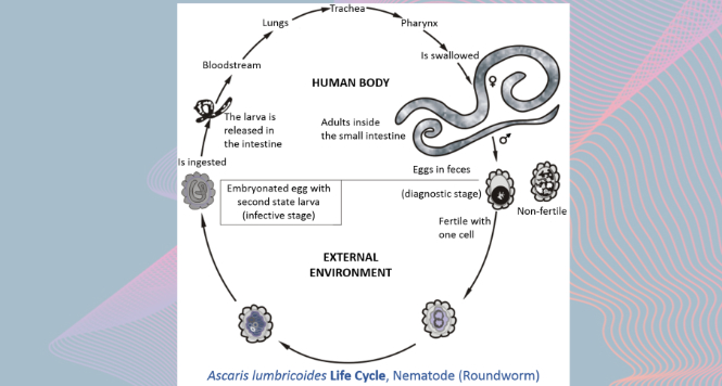
Gastric Ascaris Lumbricoides
Ascaris infestation show quite a wide array of clinical manifestations
VIEW THE GALLERY
Sub-cardial Gastric ESD
Before starting any endoscopical procedure, in particular an endoscopic submucosal
