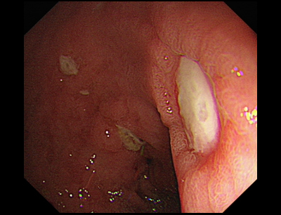
Forrest Classification
Dr. Shannon Melissa Chan
Prince of Wales Hospital, The Chinese University of Hong Kong
Forrest Classification
The Forrest Classification was first described in 1974 by J.A. Forrest et al. in TheLancet1. This classification is a widely used classification of ulcer-related upper gastrointestinal bleeding. It was initially developed to unify the description of ulcer bleeding for better communication amongst endoscopists. However, the Forrest Classification is now used as a tool to identify patients who are at an increased risk for bleeding, rebleeding and mortality2–4.
| Forrest Classification | |
| Acute haemorrhage Forrest Ia Forrest Ib | Active spurter Active oozing |
| Signs of recent haemorrhage Forrest IIa Forrest IIb Forrest IIc | Non-bleeding visible vessel Adherent clot Flat pigmented haematin on ulcer base |
| Lesions without active bleeding Forrest III | Clean-based ulcer |
Example illustrations


References:
- Forrest, JA.; Finlayson, ND.; Shearman, DJ. (Aug 1974). ‘Endoscopy in gastrointestinal bleeding’. Lancet. 2 (7877): 394–7.
- Rockall, TA, Logan, RF, Devlin, HB et al. ‘Risk assessment after acute upper gastrointestinal haemorrhage’. Gut 1996; 38: 316–21.
- Guglielmi A, Ruzzenente, A, Sandri, M et al. ‘Risk assessment and prediction of rebleeding in bleeding gastroduodenal ulcer’. Endoscopy 2002; 34: 778–86.


