Computerized Imaging of Vessels in EMR Defects Predicts Postprocedural Bleeding
Douglas K. Rex, MD, MASGE, reviewing Shaleve Y, et al. Endoscopy 2020 Sep 8.
Recent studies indicate that prophylactic clip closure of postendoscopic mucosal resection defects from colonic lesions >20 mm in size and proximal to the splenic flexure reduces the risk of delayed bleeding. However, the clipping process is expensive and time-consuming. Better prediction of bleeding could improve lesion selection for prophylactic clip closure and save money.
In a proof-of-concept study, 43 patients with clinically significant postendoscopic bleeding underwent computer-image analysis of the blood vessels in the defect. This group was compared to nonbleeding endoscopic mucosal resection (EMR) sites in 43 patients. A score based on 5 aspects of blood-vessel images yielded 86% sensitivity and 77% specificity for predicting bleeding. A neural network classifier that was built using the same independent predictors had 100% sensitivity and 77% specificity for identifying at-risk patients.

COMMENTIt is interesting that two randomized trials of cauterizing blood vessels that are visible in the base of EMR defects have not shown a benefit in preventing bleeding. However, these current data suggest that computer-based image analysis of the vessels in the defect could help select patients for prophylactic treatment of the EMR defect to prevent delayed hemorrhage.
Note to readers: At the time we reviewed this paper, its publisher noted that it was not in final form and that subsequent changes might be made.
CITATION(S)
Shaleve Y, Sabo E, Bourke MJ, Klein A. Computerized image analysis of blood vessels within mucosal defects for the prediction of delayed bleeding following colonic endoscopic mucosal resection – a pilot study. Endoscopy 2020 Sep 8. (Epub ahead of print) (https://doi.org/10.1055/a-1258-8992)
Related Posts

Polypectomy - Cold Snare
Cold snare resection has been established in diminutive polyps (up
READ THE ARTICLE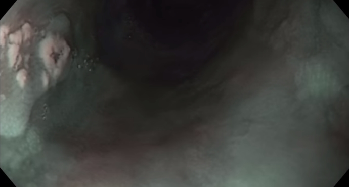
ESD Esophagus
Early esophageal carcinoma (squamous epithelium): tips and tricks for difficult
WATCH THE VIDEO