
By loading the video, you accept Vimeo's privacy policy.
Learn more
Sequenzen:
 Sequence 1: patient presentation
A 46-year-old patient with short-segment Barrett’s esophagus that had been receiving monitoring since 2009, now presenting with a mucosal adenocarcinoma.
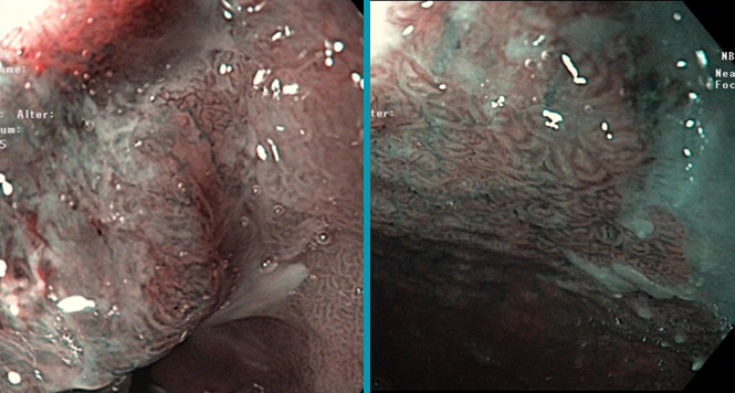
 Sequence 2: imaging of the Barrett’s segment without and with FICE
Endoscopic imaging of the short, tongue-shaped Barrett’s esophagus segment, and correlation with earlier findings before proton-pump inhibitor treatment. Above a small island of squamous epithelium at the 3-o’clock position, there is a tiny area above the esophagogastric mucosal junction from which a positive biopsy had been taken.
 Sequence 3: imaging of the lesion, with magnification
This area is imaged in detail, both in its unenhanced state and with Fujinon Intelligent Chromoendoscopy (FICE) technology, including magnification.
 Sequence 4: excision of the lesion using multiple-band ligation
Excision of the area around the small histologically positive mucosal island, using the Duette system with rubber-band ligation and loop excision.
 Sequence 5: excision surface and specimen
The excision surface, with coagulated vessel stumps. Demonstration of the mounted resection specimen. Histology: mucosal adenocarcinoma extending to the resection margin laterally.
 Sequence 6: radiofrequency therapy for the residual Barrett’s
Resection of the residual short Barrett’s segment using radiofrequency ablation (RFA). Demonstration of the whole range of accessories, from circumferential balloon ablation to focal devices, along with a focal device (TTS) that is introduced through the endoscope’s working channel. The residual Barrett’s is removed with these devices.
Related Posts
BING Classification
Multimodal therapy for early Barrett’s neoplasias, has become established as
READ THE ARTICLE
PPI, Aspirin and Prevention of Barrett’s Neoplasia
Almost everybody prescribes at least low-dose PPI to their Barrett
READ THE ARTICLE