
By loading the video, you accept Vimeo's privacy policy.
Learn more
Sequenzen:
 Sequence 1 — esophagogastroduodenoscopy (EGD) with an incidental finding of a submucosal tumor (SMT) in the mid-esophagus
A submucosal tumor was diagnosed in the mid-esophagus as an incidental finding in this patient. The lesion was progressing over time.
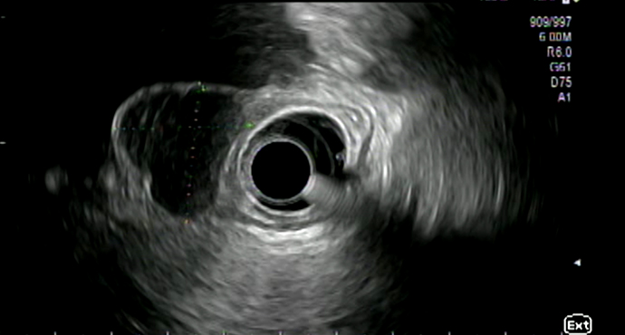
Endoscopic ultrasound (EUS) showed that the tumor was 22 mm long and 14 mm wide. Contact with the muscularis could not be definitely excluded.
 Sequence 2 — case series for SMTs in the esophagus/cardia using the tunnel technique
In this case, the indication was based on the lesion’s progression, EUS, and the patient’s desire for treatment. In view of the lesion’s size, EUS follow-up would also have been an acceptable alternative.
 Sequence 3 — endoscopic resection of the SMT using the tunnel technique: part 1, access
Submucosal injection of a methylene-stained saline solution and incision of the mucosa using the Olympus TT knife.
 Sequence 4 — endoscopic resection of the SMT using the tunnel technique: part 2, dissection of the submucosal tunnel
Dissection of the submucosal tunnel.
 Sequence 5 — endoscopic resection of the SMT using the tunnel technique: part 3, capsule-protecting tumor dissection
Capsule-protecting tumor dissection and release of the tumor from the muscularis layer.
 Sequence 6 — endoscopic resection of the SMT using the tunnel technique: part 4, mobilizing the tumor
Mobilizing the tumor out of the muscularis layer.
 Sequence 7 — endoscopic resection of the SMT using the tunnel technique: part 5, mobilizing and retrieving the tumor
At the end of the procedure, another switch is made to the Olympus IT Nano Knife to release the tumor from the muscularis.
 Sequence 8 — endoscopic resection of the SMT using the tunnel technique: part 6, resection cavity and closure of the channel
Demonstration of the excision area and closure of the mucosal entry site — histology: GIST.
Related Posts

EUS submucosal tumor in the esophagus
Submucosal lesions identified in the esophagus usually undergo further clarification
WATCH THE VIDEO