esophagus
esophagus
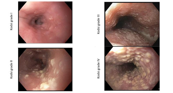
Kodsi classification of Candida esophagitis
Candidiasis is the most frequent form of infectious esophagitis. The characteristic white plaques, which are difficult to rinse off, are found in approximately 4% of
Sub-mucosal Tunneling Endoscopic Resection (STER) of an Esophageal Leiomyoma
A two centimeter submucosal lesion arising from muscularis mucosae is identified in the distal esophagus.
Neoplasia Detection Rate in Barrett’s Esophagus: A Measure of High-Quality Upper Endoscopy
Studies have reported that up to 25% of esophageal adenocarcinomas (EACs) are detected within 1 year of index endoscopy (missed cancer), emphasizing the need for
Peroral Endoscopic Myotomy Can Be Safe and Effective for Persistent Symptoms After Heller Myotomy
Persistent and recurrent symptoms have been reported in up to 20% of patients with achalasia after surgical treatment with Heller myotomy. The aim of this
Use of a Magnetically Assisted Capsule Device for Examining the Esophagus
The magnetically assisted capsule endoscopy (MACE) system (MicroCam Navi; IntroMedic) is a novel noninvasive technique for visualizing the upper GI tract. This device utilizes a
Patients With Barrett’s Esophagus and Indefinite Dysplasia: What To Do?
Prateek Sharma, MD, FASGE, reviewing Krishnamoorthi R, et al. Gastrointest Endosc 2020 Jan. The risk of progression in Barrett’s esophagus (BE) with low-grade dysplasia has

Achalasia: Chicago Classification
Achalasia is one of the differential diagnoses in patients with symptoms of dysphagia. High-resolution (HR) manometry is now regarded as the diagnostic gold standard for

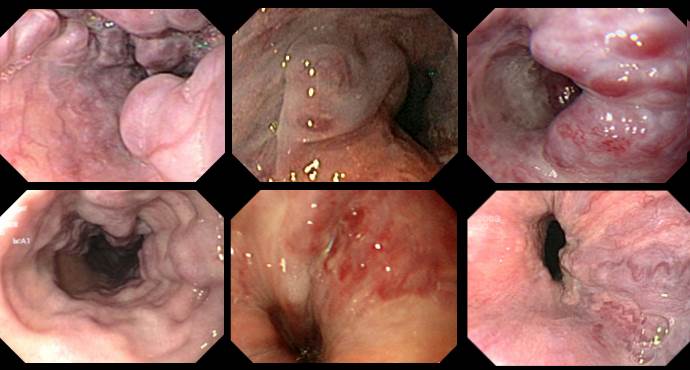
Paris Classification: Early Squamous Cell Cancers Esophagus
Examples of superficial/early squamous cell lesions in the esophagus are presented below. In the esophagus, flat lesions are predominant in the early tumors, and polypoid

Heterotopic gastric mucosa
Heterotope Magenschleimhaut des Ösophagus (heterotopic gastric mucosa, gastric inlet patch) entspricht funktionellem Magengewebe, das sich nicht an der anatomisch üblichen Lokalisation befindet. Sie ist in
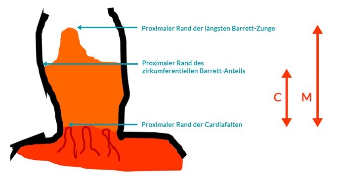
Prague Classification Barrett Esophagus
The Prague classification was presented by an international research group in 2006 (1) and has since been regarded as the standard for measuring the length

Reflux Esophagitis: Los Angeles Classification
Gastroesophageal reflux disease with endoscopically identifiable lesions (erosions, stricture, Barrett’s esophagus) is defined as erosive gastroesophageal reflux disease (GERD). Fewer than 50% of patients with
Esophageal Varices
Various systems are available for classifying esophageal varices. Unfortunately, they only overlap or coincide partly. The official terminology used by the German Society for Digestive

Achalasia — place of endoscopic therapy in the light of the first long-term data for POEM
Achalasia is a rare neuromuscular disease of unclear etiology that probably has a genetic background. The precise etiopathogenesis of achalasia is still unclear. Above all,
